U.C.L.A. Rheumatology Pathophysiology of Disease Course Lecture,
Second Year Medical School 1997


  FirstPreviousNextLast Page 14


Rheumatoid Arthritis     Page 14
  ......Index Go to Previous page .....Go to Next page


      damage. They are the predominant lymphocyte population in the proliferative synovium and they release cytokines which amplify and perpetuate inflammation in the joints and activate B cells (figure 5). They are believed to be activated through the RA-related MHC Class II epitope described above in combination with a disease-provoking antigenic peptide or with a super-antigen that binds to the MHC. The pathogenic sets of T cells are suspected to share certain common T cell receptors.
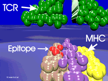
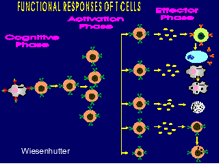
   
Figure 4. A closeup of the schematic drawing of HLA-DR4 in figure 3. Click thumbnail figure to view full graphic. 219 x 164 pixels 16kbs
 
Click Picture or here for larger slide and further discussion (640 x 480) 53kb
  FirstPreviousNextLast
Figure 5. A scheme demonstrating the effects of T cell activation is depicted. Click thumbnail figure to view full graphic.. 219 x 164 pixels 10kbs
Click Picture or here for larger slide and further discussion (640 x 480) 44kb
  Page 14
  ......Index Go to Previous page .....Go to Next page
About Us | Contact Us | ©2005 This web site has been developed and maintained by Craig W. Wiesenhutter, M.D.