U.C.L.A. Rheumatology Pathophysiology of Disease Course Lecture, Second Year Medical School 2005 |
|||||||||||||||||||||||||||||||||||
Page 56 | |||||||||||||||||||||||||||||||||||
......Index Go to Previous page .....Go to Next page | |||||||||||||||||||||||||||||||||||
Pathophysiology of Spondyloarthropathies: Theory #6 HLA-B27 Causes Arthritis By Being Degraded, and then Provides a Peptide to be Presented by HLA Class II molecules Page 56 |
|||||||||||||||||||||||||||||||||||
Windows Media Player |
QuickTime Media Player |
Real Media Player |
|||||||||||||||||||||||||||||||||
A degraded HLA-B27 molecule is shown being presented to a helper T-cell. |
Animation: SpA Pathophysiology #6: A degraded HLA-B27 molecule is shown being presented to a helper T-cell. |
Animation: SpA Pathophtysiology #6: A degraded HLA-B27 molecule is shown being presented to a helper T-cell. |
||||||||||||||||||||||||||||||||
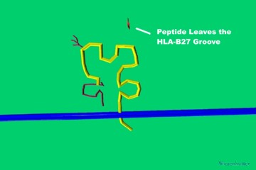
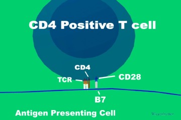
Slide 1: "The Intact HLA-B27 Molecule is shown on the Cell Surface of an Antigen Presenting Cell " 360 x 240 pixels 12kb drawn freehand in 3dStudio Max.x. | Slide 2: "The Peptide is lost from the HLA-B27's Groove " 360 x 240 pixels 11kb drawn freehand in 3dStudio Max. | Slide 3: "The loss of the Peptide from the HLA-B27 Groove results in Molecular Instability " 360 x 240 pixels 12kb drawn freehand in 3dStudio Max. | |||||||||||||||||||||||||||||||||
Click Picture or here for larger slides (720 x 480 ) 31kb | Click Picture or here for larger slides (720 x 480 ) 27kb | Click Picture or here for larger slides (800 x 500 ) 31kb | |||||||||||||||||||||||||||||||||
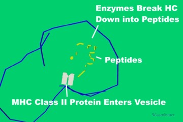
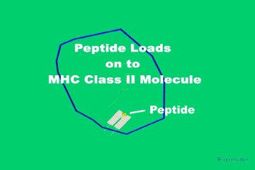
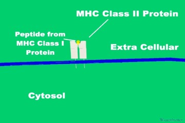
The presence of such findings radically alters the classical views of pathogenesis. There is a possibility that HLA-B27 might cause arthritis by being degraded, and then providing a peptide to be presented by HLA class II molecules, as shown here. If such were the case, then the mediators of arthritis would be CD4 positive T cells. |
|||||||||||||||||||||||||||||||||||
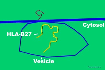
Slide 4: "The HLA-B27 Heavy Chain is taken up by an Intracellular Vesicle " 360 x 240 pixels 14kb drawn freehand in 3dStudio Max. | Slide 5: "The HLA-B27 Heavy Chain is broken down into Peptides: a MHC Class II Protein Enters the Vesicle " 360 x 240 pixels 17kb drawn freehand in 3dStudio Max | ||||||||||||||||||||||||||||||||||
Slide 6: "A Peptide from the HLA-B27 Heavy Chain Loads on to the MHC Class II Molecule" 360 x 240 pixels 12kb drawn freehand in 3dStudio Max. | |||||||||||||||||||||||||||||||||||
Click Picture or here for larger slides (720 x 480 ) 35kb | Click Picture or here for larger slides (720 x 480 ) 44kb | Click Picture or here for larger slides (720 x 480 ) 45kb | |||||||||||||||||||||||||||||||||
Slide 7: "The Peptide from the HLA-B27 Heavy Chain is Displayed by the MHC Class II Molecule on the Cell surface of the Antigen Presenting Cell " 360 x 240 pixels 13kb drawn freehand in 3dStudio Max. | Slide 8: "A CD4 Positive T-Cell Interacts with the Antigen Presenting Cell: Both of the necessary Signals for T-Cell activation will occur as all the required Receptors are present, as shown " 360 x 240 pixels 15kb drawn freehand in 3dStudio Max. | ||||||||||||||||||||||||||||||||||
Page 56 | |||||||||||||||||||||||||||||||||||
Click Picture or here for larger slides (720 x 480 ) 33kb | Click Picture or here for larger slides (720 x 480 ) 37kb | ||||||||||||||||||||||||||||||||||
......Index Go to Previous page .....Go to Next page | |||||||||||||||||||||||||||||||||||
This web site has been developed and maintained by Craig W. Wiesenhutter, M.D. | |||||||||||||||||||||||||||||||||||
About Us | Contact Us | ©2005 | |||||||||||||||||||||||||||||||||||