The Cytotoxic T-Cell Response, Cloned Armed T-Cells, First and Second T-Cell Activation Signals, and Apoptosis Part II Page 51
Windows Media Player
QuickTime Media Player
Real Media Player
C
loned and armed T-cells reenter the circulation and then migrate from the vessel to the site of inflammation.
Animation: Cytotoxicity #2 : Cloned and armed T-cells reenter the circulation and then migrate from the vessel to the site of inflammation.
Animation: Cytotoxicity #2 : Cloned and armed T-cells reenter the circulation and then migrate from the vessel to the site of inflammation.
Animation: Cytotoxicity #2 : Cloned and armed T-cells reenter the circulation and then migrate from the vessel to the site of inflammation.
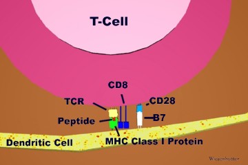
Slide 1: "A peptide MHC trimolecular complex, TCR, and the receptors responsible for the second signal, B7 & CD28, are Labeled" 360 x 240 pixels 17kb drawn freehand in 3dStudio Max.
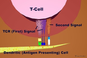
Slide 2: "The TCR (First) Signal and the Second Signal, within the T-Cell, is illustrated " 360 x 240 pixels 18kb drawn freehand in 3dStudio Max.
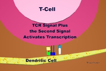
Slide 3: "TCR Signal Plus the Second Signal Activates Transcription " 360 x 240 pixels 17kb drawn freehand in 3dStudio Max.
When a peptide MHC trimolecular complex is recognized by the TCR, and the appropriate co-stimulatory signals are also generated, the transduced signals will ultimately lead to T cell activation, proliferation, and maturation.
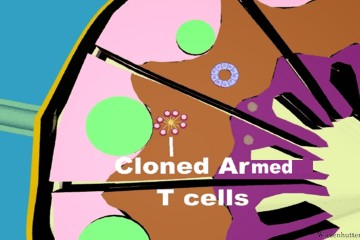
Slide 4: "After several days, Cloned Armed T-Cells develop, as drawn here " 360 x 240 pixels 25kb drawn freehand in 3dStudio Max..
Slide 5: "Armed T-Cells leave the Lymph Node via the Efferent Lymphatic Vessel " 360 x 240 pixels 30kb drawn freehand in 3dStudio Max.
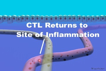
Slide 6: "A Cytotoxic T Lymphoctye is seen migrating, via a blood vessel, to the site of Inflammation " 360 x 240 pixels 18kb drawn freehand in 3dStudio Max.
Over a period of several days, these cloned T cells multiply and become fully armed. Once armed, they also leave the lymph node, and reenter the blood stream via the thoracic duct.
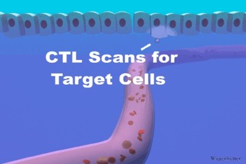
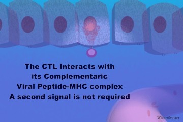
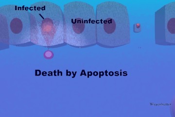
The armed CTL home to the site of infection by means of new adhesion molecules and chemotaxic factors induced by the inflammatory response. A CTL is seen extravasating from a blood vessel and then scanning epithelial cells for its specific peptide MHC Class I complex, in this case a viral peptide. When a complex is recognized, the CTL rapidly and efficiently kills the infected cell by apoptosis, and then moves on to scan more cells. Co-stimulatory signals are not required.
Slide 7: "A Cytotoxic T Lymphoctye scans Target Cells " 360 x 240 pixels 15kb drawn freehand in 3dStudio Max
Slide 8: "The CTL Interacts with its Complementaric Viral Peptide-MHC complex. A second signal is not required " 360 x 240 pixels 19kb drawn freehand in 3dStudio Max
Slide 9: "Death by Apoptosis is Displayed #1" 360 x 240 pixels 14kb drawn freehand in 3dStudio Max
Slide 10: "Death by Apoptosis is Displayed #2" 360 x 240 pixels 14kb drawn freehand in 3dStudio Max