U.C.L.A. Rheumatology Pathophysiology of Disease Course Lecture, Second Year Medical School 2005


      Page 49
 
......Index Go to Previous page .....Go to Next page
 
 
Translation of Membrane Bound Proteins such as MHC Class I Proteins, the Golgi Apparatus, and the Plasma Membrane  Part II  Page 49
 
 
Windows Media Player
QuickTime Media Player
Real Media Player
 

The translation of MHC class I protein starts at the cytosol ER border and is followed as it is folded and interacts with the TAP complex and then the molecule is further followed from the TAP complex until it has traveled to and is displayed on the cell surface.

Animation: Translation #3
: The molecule is further followed from the TAP complex until it has traveled and is displayed on the cell surface.

Animation: Translation #3
: The molecule is further followed from the TAP complex until it has traveled and is displayed on the cell surface
Animation: Translation #3
: The molecule is further followed from the TAP complex until it has traveled and is displayed on the cell surface
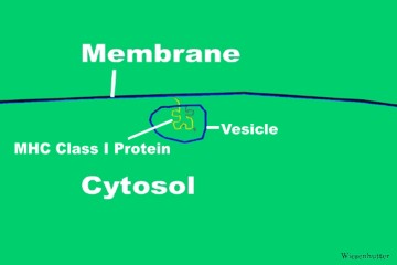
 
       
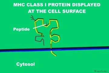
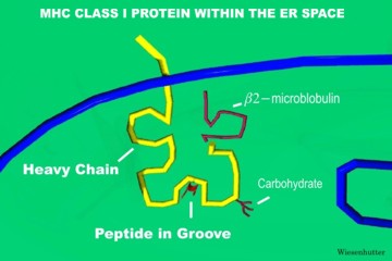
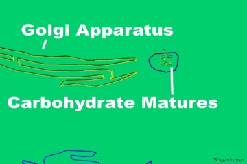
Slide 1: "The MHC Class I Protein is drawn within the ER Space " 360 x 240 pixels 19kb drawn freehand in 3dStudio Max. Slide 2: "The MHC Class I's Carbohydrate "Matures" as the Vesicle nears the Golgi Apparatus " 360 x 240 pixels 15kb drawn freehand in 3dStudio Max. Slide 3: "The MHC Class I Protein approaches the Plasma Membrane within its Vesicle " 360 x 240 pixels 11kb drawn freehand in 3dStudio Max
Click Picture or here for larger slides (720 x 480 ) 50kb Click Picture or here for larger slides (720 x 480 ) 49kb Click Picture or here for larger slides (720 x 480 ) 40kb
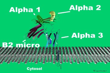
            The MHC Class I molecule makes its way to the membrane surface via a vesicle. The carbohydrate maturates in the Golgi apparatus. The MHC Class I protein at the end of the animation is shown as a ribbon structure, revealing its secondary structure of alpha helix and beta pleated sheets.


Slide 4: "The MHC Class I Protein is Displayed at the Cell Surface " 360 x 240 pixels 16kb drawn freehand in 3dStudio Max. Slide 5: "The Ribbon Structure of a MHC Class I in the Plasma Membrane replaces the previous schematic drawing " 360 x 240 pixels 27kb drawn freehand in 3dStudio Max.
Click Picture or here for larger slides (720 x480 ) 29kb Click Picture or here for larger slides (800 x 600 ) 87kb
  Page 49
     
......Index Go to Previous page .....Go to Next page
This web site has been developed and maintained by Craig W. Wiesenhutter, M.D.
About Us | Contact Us | ©2005