U.C.L.A. Rheumatology Pathophysiology of Disease Course Lecture,
Second Year Medical School 1997
Rheumatoid Arthritis Figure 3

U.C.L.A. Rheumatology Pathophysiology of Disease Course Lecture, |
|||||||||||||||
Rheumatoid Arthritis Figure 3 |
Page 48 | ||||||||||||||
 |
|||||||||||||||
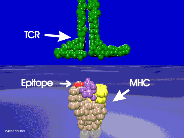
| Figure 3. A schematic drawing of HLA-DR4 (MHC or major histocompatability complex) is depicted in the surface of an antigen presenting cell. The alpha chain is shown in yellow and the beta chain is shown in beige. The antigenic peptide is shown in purple and is nestled in the antigenic cleft surrounded on either side by alpha helixes of the HLA molecule. The disease associated epitope is shown in red. Note the location in the alpha helix of the beta chain. These amino acids are situated in such a way as to be associated with or in direct contact to either the antigenic peptide or the T cell receptor (TCR) or both.640 x 480 pixels 61kbs freehand 3dstudio max | |||||||||||||||
| Click Picture or here to Return | |||||||||||||||
| Page 48 | |||||||||||||||
| About Us | Contact Us | ©2005 | This web site has been developed and maintained by Craig W. Wiesenhutter, M.D. | ||||||||||||||