U.C.L.A. Rheumatology Pathophysiology of Disease Course Lecture, Second Year Medical School 2005
The structures of the normal joint are described.
Animation: Normal Joint Anatomy : The structures of the normal joint are described.
Animation: Normal Joint Anatomy : The structures of the normal joint are described.
Animation: Normal Joint Anatomy : The structures of the normal joint are described.


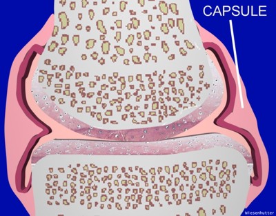
The joint capsule is a fibrous structure that defines the outer boundary of joints. The thickness of the capsule varies widely depending on the type of joint and the location within a given joint. For instance, a thickened capsule prevents hyperextension in most hinge joints. Parallel collagen fibers, known as ligaments, run within the capsule and contributes to maintaining the joint's integrity.
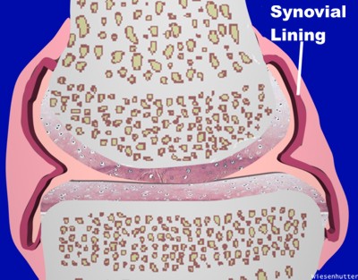
A thin lining of cells, one to three cells thick, runs beneath the capsule and is called the synovium. The synovium is comprised of two types of cells: Type I cells, that are histologically similar to macrophages, and Type II cells, which resemble fibrocytes. This lining of cells produces synovial fluid that enters the joint space and acts as a lubricant. It also stabilizes the joint by acting as an adhesive.
Synovial joints are characterized by the presence of hyaline cartilage, the composition of which results in a remarkably long lasting load bearing surface. This structure, working in concert with synovial fluid, results in near frictionless movement.

