U.C.L.A. Rheumatology Pathophysiology of Disease Course Lecture, Second Year Medical School 2005 |
||||||||||||||||||||||||||||||||||||||||
Page 46 | ||||||||||||||||||||||||||||||||||||||||
......Index Go to Previous page .....Go to Next page | ||||||||||||||||||||||||||||||||||||||||
Adenoviral (Adenovirus) and Chlamydial Infection, and a HLA-B27 Positive Patient, a Dendritic cell and a Macrophage Page 46 |
||||||||||||||||||||||||||||||||||||||||
Windows Media Player |
QuickTime Media Player |
Real Media Player |
||||||||||||||||||||||||||||||||||||||
The immune response to infection by the adenovirus in the lung, and chlamydia in the genitourinary system is animated. |
Animation: Chlamydial Infection #1 : The immune response to infection by the adenovirus in the lung, and chlamydia in the genitourinary system is animated Animation: Chlamydial Infection #2. |
Animation: Chlamydial Infection #1 : The immune response to infection by the adenovirus in the lung, and chlamydia in the genitourinary system is animated. Animation: Chlamydial Infection #2 |
Animation: Chlamydial Infection #1: The immune response to infection by the adenovirus in the lung, and chlamydia in the genitourinary system is animated. Animation: Chlamydial Infection #2 |
|||||||||||||||||||||||||||||||||||||
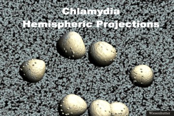
Slide 1: "The vegetative form of Chlamydia is shown here, the Elementary Body (EB)" 360 x 240 pixels 17kb drawn freehand in 3dStudio Max. Bitmap made in CorelDraw | Slide 2: "This is a Rendition of an EM showing the Hemispheric Projections of the elemental bodies" 360 x 240 pixels 51kb drawn freehand in 3dStudio Max. |
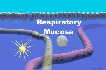
Slide 3: "The good Doctor's Respiratory Tract Mucosa " 360 x 240 pixels 19kb drawn freehand in 3dStudio Max. |
||||||||||||||||||||||||||||||||||||||
Click Picture or here for larger slides (720 x 480 ) 45kb | Click Picture or here for larger slides (720 x 480 ) 179kb | Click Picture or here for larger slides (720 x 480 ) 55kb | ||||||||||||||||||||||||||||||||||||||
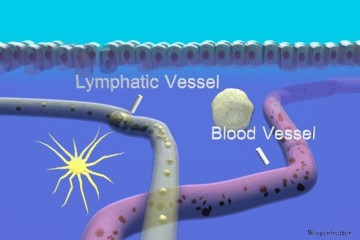
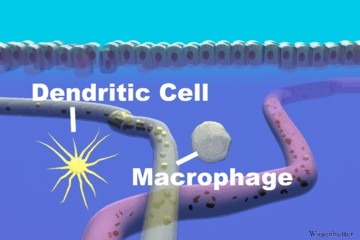
Chlamydia is an obligate intracellular bacterium. It has two forms, including the vegetative form shown here, the elementary body (EB). The double layered membrane is rigid and relatively impermeable. The EB's metabolism is not active and this form of the organism can withstand the rigors of the extracellular environment. This is a rendition of an EM showing the hemispheric projections of the elemental bodies. Their purpose is not known. This scene represents the initial immunologic battlefield, the sub mucosa of the Doctor's respiratory tract and Chubs' genitourinary tract. A macrophage is seen on patrol, while a dendritic cell awaits at its station, sampling extracellular materials for antigens by the means of macropinocytosis. A blood vessel and lymph vessel are shown.”. |
||||||||||||||||||||||||||||||||||||||||
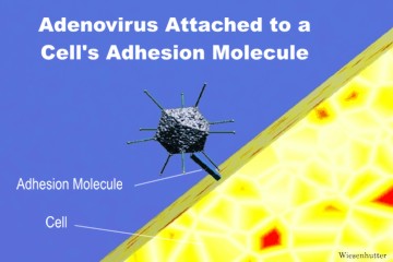
Slide 4: "A Dendritic Cell and a Macrophage Patrols the Submucosa " 360 x 240 pixels 20kb drawn freehand in 3dStudio Max. | Slide 5: "The Vessels of the Respiratory Submucosa are depicted " 360 x 240 pixels 19kb drawn freehand in 3dStudio Max. | Slide 6: "An Adenovirus Attaches to a Human Cell's Adhesion Molecule " 360 x 240 pixels 20kb drawn freehand in 3dStudio Max | ||||||||||||||||||||||||||||||||||||||
Click Picture or here for larger slides (720 x 480 ) 55kb | Click Picture or here for larger slides (720 x 480 ) 56kb | Click Picture or here for larger slides (720 x 480 ) 57kb | ||||||||||||||||||||||||||||||||||||||
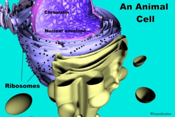
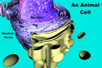
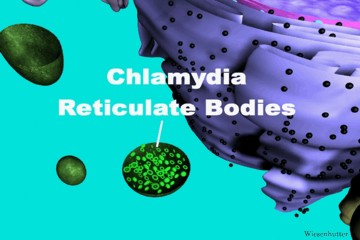
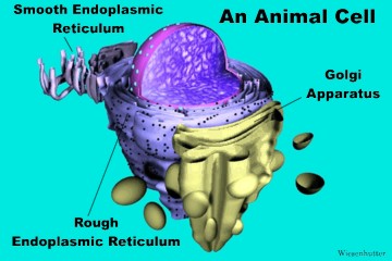
Both Chlamydia and Adenovirus readily adhere to cells. Chlamydia can even induce phagocytosis in nonphagocytic cells. An Adenovirus is depicted initially adhering, through its fibers, to an adhesion molecule on the dendritic cell. The virus is internalized in endosomes and undergoes an uncoating. The sub cellular structures that are important to the discussion are shown. The endomembrane system consists of the Golgi apparatus, and rough and smooth endoplasmic reticulum. The nucleus and nuclear pores are depicted. Note that the double layered outer membrane is continuous with the endomembrane system, and that the nuclear pores are continuous with the cytosol. Once the Chlamydia enters the phagosomes of the cell, there is a morphologic change to the reticulate bodies (RB), as shown here. In this stage the organism is metabolically active and they divide, but they are quite fragile. Lysosomes are inhibited from fusing with the phagosomes until just before host cell death. |
||||||||||||||||||||||||||||||||||||||||
Slide 7: "An Animal Cell is drawn: Important Organelles are delineated #1" 360 x 240 pixels 28kb drawn freehand in 3dStudio Max | ||||||||||||||||||||||||||||||||||||||||
Click Picture or here for larger slides (720 x 480 ) 80kb | ||||||||||||||||||||||||||||||||||||||||
Slide 8: "An Animal Cell is drawn: Important Organelles are delineated #2" 360 x 240 pixels 28kb drawn freehand in 3dStudio Max | Slide 9: "An Animal Cell is drawn: Important Organelles are delineated #3" 360 x 240 pixels 29kb drawn freehand in 3dStudio Max | Slide 10: "A Chlamydia in a Human Cell Phagosome is drawn: there is a Morphologic change to the Reticulate Bodies (RB), as shown here." 360 x 240 pixels 25kb drawn freehand in 3dStudio Max | ||||||||||||||||||||||||||||||||||||||
Click Picture or here for larger slides (720 x 480 ) 80kb | Click Picture or here for larger slides (720 x 480 ) 85kb | Click Picture or here for larger slides (720 x 480 ) 69kb | ||||||||||||||||||||||||||||||||||||||
Page 46 | ||||||||||||||||||||||||||||||||||||||||
......Index Go to Previous page .....Go to Next page | ||||||||||||||||||||||||||||||||||||||||
About Us | Contact Us | ©2005 | This web site has been developed and maintained by Craig W. Wiesenhutter, M.D. | |||||||||||||||||||||||||||||||||||||||