U.C.L.A. Rheumatology Pathophysiology of Disease Course Lecture, Second Year Medical School 2005 |
||||||||||||||||||||||||||||||||||||||||||
Page 29 | ||||||||||||||||||||||||||||||||||||||||||
......Index Go to Previous page .....Go to Next page | ||||||||||||||||||||||||||||||||||||||||||
Rheumatoid Arthritis, Characteristic Histologic Changes Page 29 |
||||||||||||||||||||||||||||||||||||||||||
Windows Media Player |
QuickTime Media Player |
Real Media Player |
||||||||||||||||||||||||||||||||||||||||
Examples of histopathology, typical xrays, and photos of ra hands are shown. |
Animation: Rheumatoid Arthritis and Joint Changes#1 : The typical joint changes in RA are described. Animation: Rheumatoid Arthritis and Joint Changes#2 : Examples of histopathology, typical xrays, and photos of ra hands are shown. |
Animation: Rheumatoid Arthritis and Joint Changes#1: The typical joint changes in RA are described. Animation: Rheumatoid Arthritis and Joint Changes#2 : Examples of histopathology, typical xrays, and photos of ra hands are shown. |
|||||||||||||||||||||||||||||||||||||||
Slide 1: "Normal Joint Space is Shown: The Synovial Lining is Just a few cells thick " 400 x 300 pixels 23kb freehand in 3dStudio Max | Slide 2: "The Rheumatoid Arthritis Joint Space is Shown " 400 x 300 pixels 36kb freehand in 3dStudio Max | |||||||||||||||||||||||||||||||||||||||||
Click Picture or here for larger slides (800 x 600 ) 66kb | Click Picture or here for larger slides (800 x 600 ) 109kb | |||||||||||||||||||||||||||||||||||||||||
Let us return to our discussion of inflammation, and the immune response. I would now like to discuss RA further. The synovial lining and subsynovial space in the normal joint is shown. In RA a number of changes to the joint space takes place, including thickening of the synovial lining from just a few cells thick to many cells. And there is the formation of new blood vessels and a marked infiltration of mononuclear cells into the region, as shown here. Also the Rheumatoid joint is known to contain substantial quantities of immunoglobulin, formulated as aggregates, in the synovium, synovial fluid, cartilage and fibrocartilage. The accumulation of aggregates within the rheumatoid joint has been considered important in the pathogenesis of RA. We will return to this topic shortly. |
||||||||||||||||||||||||||||||||||||||||||
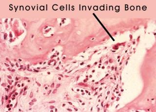
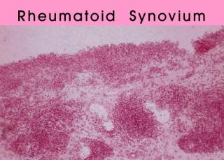
Slide 3: "Normal synovium."The synovial lining cells are only a few cells thick. From Dieppe: textbook of rheumatology. | Slide 4: "Rheumatoid Synovium. "Marked infiltration of mononuclear cells. From Dieppe: textbook of rheumatology. | Slide 5: "Rheumatoid Synovium. Synovial cells are invading bone. From Dieppe: textbook Rheumatology." | ||||||||||||||||||||||||||||||||||||||||
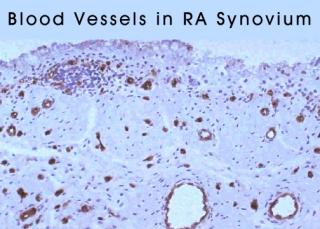
We know take closer look at the subsynovial space in a rheumatoid joint. A histological sample of a normal synovial lining is shown, followed by a synovial biopsy from a patient with RA. The mononuclear infiltrate is evident. This image shows RA tissue stained for blood vessels, demonstrating their increase. Synovial tissue is shown here invading bone. Such destructive behavior by the synovial cells leads to the characteristic x-ray findings of marginal erosions as shown here. In aggregate, the destructive nature of the disease process can lead to progressive deformity and startling crippling. This patients hands were photographed in 1992, and then again ten years later, as shown here. |
||||||||||||||||||||||||||||||||||||||||||
Slide 6: "Rheumatoid Synovium." There is marked angiogenesis. From Dieppe: textbook of rheumatology. | Slide 7: "The characteristic x-ray findings of marginal erosions is shown " 300 x 225 jpeg 22kb CAC | Slide 8: "A photo of a ra patient's hands taken in 1992 " 300x 200 13kb CAC | ||||||||||||||||||||||||||||||||||||||||
Click Picture or here for larger slides (720 x 480 ) 101kb | Click Picture or here for larger slides (720 x 480 ) 72kb | |||||||||||||||||||||||||||||||||||||||||
Slide 5: "The same Patient with Rheumatoid Arthritis Hands in 2002 " 400 x 300 pixels 24kb photo CAC | Slide 6: "The Patient with Rheumatoid Arthritis Hands are Compared 1992 and 2002 " 400 x 300 pixels 26kb photo CAC | |||||||||||||||||||||||||||||||||||||||||
Click Picture or here for larger slides (800 x 600 ) 58kb | Click Picture or here for larger slides (800 x 600 ) 58kb | |||||||||||||||||||||||||||||||||||||||||
Page 29 | ||||||||||||||||||||||||||||||||||||||||||
......Index Go to Previous page .....Go to Next page | ||||||||||||||||||||||||||||||||||||||||||
About Us | Contact Us | ©2005 | This web site has been developed and maintained by Craig W. Wiesenhutter, M.D. | |||||||||||||||||||||||||||||||||||||||||