U.C.L.A. Rheumatology Pathophysiology of Disease Course Lecture, Second Year Medical School 2005 |
||||||||||||||||||||||||||||||
Page 27 | ||||||||||||||||||||||||||||||
......Index Go to Previous page .....Go to Next page | ||||||||||||||||||||||||||||||
Systemic Lupus Erythematosus Inflammation and Immune Complex Disease Page 27 |
||||||||||||||||||||||||||||||
Windows Media Player |
QuickTime Media Player |
Real Media Player |
||||||||||||||||||||||||||||
SLE is primarily an immune complex disease. The effects of an immune complex forming on a bacteria is animated. |
Animation: SLE : Immune injury in SLE is illustrated. Animation: SLE #2: Immune Complex Deposition : SLE is primarily an immune complex disease. The effects of an immune complex forming on a bacteria is animated. |
|||||||||||||||||||||||||||
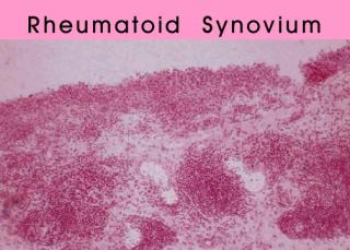
Slide 1: " Rheumatoid Synovium " 320 x 229 pixels 17kb from Dieppe Textbook of Medicine | ||||||||||||||||||||||||||||||
Slide 2: " Synovitis in a Patient with Systemic Lupus Erythematosus " 400 x 280 pixels 25kb photo CAC Worked (animated) in 3dStudio Max | ||||||||||||||||||||||||||||||
Click Picture or here for larger slides (800 x 561 ) 82kb | ||||||||||||||||||||||||||||||
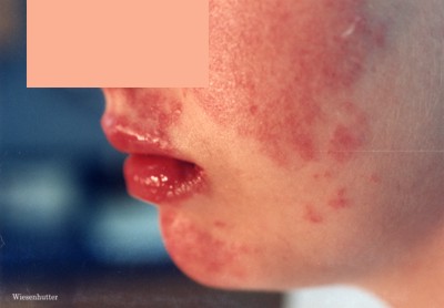
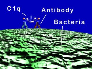
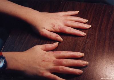
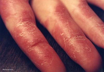
The accumulation of inflammatory cells in a rheumatoid joint is called synovitis. This young lady also had synovitis in a rheumatoid pattern, but there were several other finding as well. The rash is noted, and extended to the volar surface of the hand as shown. The rash also involved her face. A urinalysis revealed active kidney injury. The pattern of both joint and organ involvement leads to a diagnosis of SLE. SLE is an immune complex disease. Again, in most situations, the formation of immune complexes is a good thing. The power of combining an immune complex with the destructive force of complement is shown in this cartoon. Two antibodies have combined with the cell surface of a bacterium. The complement component, C1Q reacts with the Fc portion of the two antibody molecules, initiating the complement cascade. The inflammatory response is enhanced by the formation of small complement fragments, which will direct white cells to the site, in a process called chemotaxis. The membrane attack complex is formed, instills itself into the bacterium's membrane, leading to osmotic destruction of the cell. Here, this immune response is beneficial to the body, but imagine such destructive force directed at the human kidney, as with the patient shown above. |
||||||||||||||||||||||||||||||
Slide 4: "Facial Rash in a Patient with Systemic Lupus Erythematosus " 400 x 278 pixels 19kb photo CAC Worked (animated) in 3dStudio Max | ||||||||||||||||||||||||||||||
Slide 3: " Volar Surface of the hand in a Patient with Systemic Lupus Erythematosus " 400 x 280 pixels 26kb photo CAC Worked (animated) in 3dStudio Max | ||||||||||||||||||||||||||||||
Click Picture or here for larger slides (800 x 559) 83kb | Click Picture or here for larger slides (800 x 600 ) 38kb | |||||||||||||||||||||||||||||
Page 27 | ||||||||||||||||||||||||||||||
Slide 5: " An Immune Complex forms on the Surface of a Bacterium " 320 x 240 pixels 27kb drawn freehand in 3dStudio Max | ......Index Go to Previous page .....Go to Next page | |||||||||||||||||||||||||||||
About Us | Contact Us | ©2005 | This web site has been developed and maintained by Craig W. Wiesenhutter, M.D. | |||||||||||||||||||||||||||||