U.C.L.A. Rheumatology Pathophysiology of Disease Course Lecture, Second Year Medical School 2005
The Collagen Molecule, Aggrecan, Hyaluronate and Osteogenesis Imperfecta
The structure of collagen is described.
Animation: Collagen Structure : The structure of collagen is described.
Animation: Collagen Structure : The structure of collagen is described.
Animation: Collagen Structure : The structure of collagen is described.
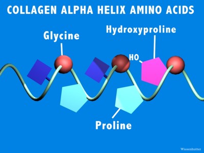
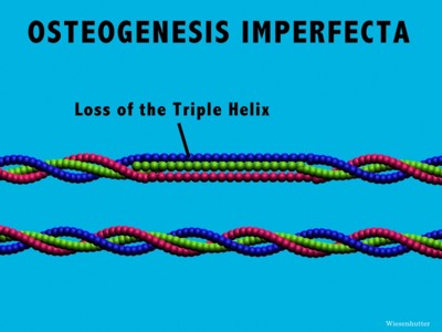
There are several different kinds of collagen, more than 14 have been described so far, which are defined by their unique series of amino acids. The chemical structure of these amino acids encourage the formation of the triple helix. Type I and Type II collagen are the most important collagens in the matrix of bone and cartilage, respectively.
Osteogenesis Imperfecta is a disease in which there is a substitution of a cysteine for the glycine of the collagen helix, which then sterically hinders the formation of the triple helix.
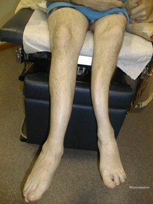
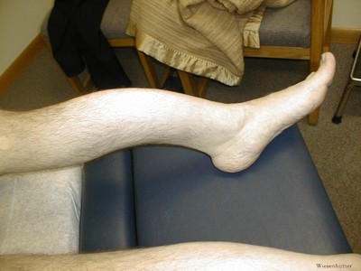
This genetic disease results in a dramatic weakening in the structure of bone, which leads to frequent fractures occurring at an early age. The fractures, in turn, lead to permanent deformaties of the extremities, as shown with this patient.
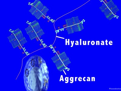
In addition to collagen, the other major constituent of the cartilage matrix is proteoglycans. The most important proteoglycan is aggrecan. Aggrecan is a giant, complex macromolecule. The polysaccharide side chains are hydrophilic and attract water. They also have multiple negative charges, which cause the molecules to repel each other.
The aggrecan is then attached to a hyaluronate core molecule.
