U.C.L.A. Rheumatology Pathophysiology of Disease Course Lecture,
Second Year Medical School 1997
Rheumatoid Arthritis Figure 6

U.C.L.A. Rheumatology Pathophysiology of Disease Course Lecture, |
|||||||||||||||
Rheumatoid Arthritis Figure 6 |
Page 51 | ||||||||||||||
 |
|||||||||||||||
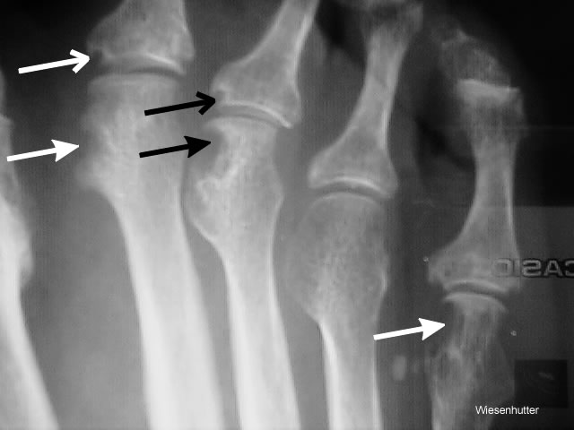
| Figure 6. A foot xray from a 25 year old women with RA clearly shows erosive bone damage. The large headed arrows show large erosions whereas the small headed arrows show smaller marginal erosions (mouse ear erosion). Deformity, as reflected by the loss of the normal straight alignment of the bones, is already evident in this young woman. jpeg 640 x 480 pixels 30kbs photo CAC | |||||||||||||||
| Click Picture or here to Return | |||||||||||||||
| Page 51 | |||||||||||||||
| About Us | Contact Us | ©2005 | This web site has been developed and maintained by Craig W. Wiesenhutter, M.D. | ||||||||||||||