U.C.L.A. Rheumatology Pathophysiology of Disease Course Lecture,
Second Year Medical School 1997
Introduction Figure 5
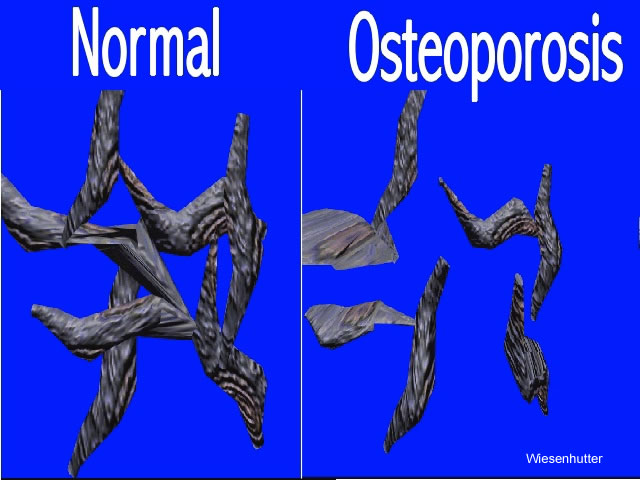
Figure 5. A schematic representation of normal trabeculae of bone (left) vs the trabeculae in bone with osteoporosis is shown. Note that there is not only a reduction of the size of the trabeculae (bone mass reduction) but that there is also a loss in the number of bony connections and bridges (microarchitectual defect). Whereas both of these processes lead to bony fragility, only bone mass is assessed by the commonly employed diagnostic tests (DEXA, QCT). Never-the-less, the tests for diagnosing osteoporosis represent some of the most accurate tests for diagnoses in medicine today. jpeg 640 x 480 pixels 57kbs