RHEUMATOID ARTHRITIS: SLIDES & ANIMATIONS
PAGE 4
RA is a common severe inflammatory disorder and is marked by a variable course. The disease is multi factorial in origin and includes a genetic predisposition. It is a multisystem disease, but we will focus on the hallmark of the disorder, i.e. its attack on joints (synovitis).
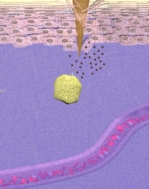
The synovial lining and subsynovial space in the normal joint is shown. In RA a number of changes to the joint space takes place, including thickening of the synovial lining from just a few cells thick to many cells. And there is the formation of new blood vessels and a marked infiltration of mononuclear cells into the region, as shown here. Also the Rheumatoid joint is known to contain substantial quantities of immunoglobulin, formulated as aggregates, in the synovium, synovial fluid, cartilage and fibrocartilage. The accumulation of aggregates within the rheumatoid joint has been considered important in the pathogenesis of RA. We will return to this topic shortly.


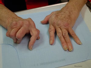
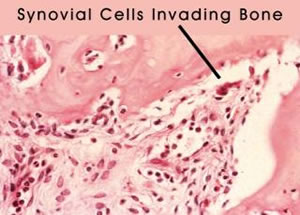
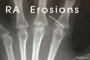
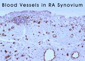
A histological sample of a normal synovial lining is shown, followed by a synovial biopsy from a patient with RA. The mononuclear infiltrate is evident. This image shows RA tissue stained for blood vessels, demonstrating their increase. Synovial tissue is shown here invading bone. Such destructive behavior by the synovial cells leads to the characteristic x-ray findings of marginal erosions as shown here. In aggregate, the destructive nature of the disease process can lead to progressive deformity and startling crippling. This patient’s hands were photographed in 1992, and then again ten years late.
Animation 9b: Rheumatoid Arthritis and Joint Changes#2 : Examples of histopathology, typical xrays, and photos of ra hands are shown.
Animation9a: Rheumatoid Arthritis and Joint Changes#1 : The typical joint changes in RA are described.
Animation 9b: Rheumatoid Arthritis and Joint Changes#2 : Examples of histopathology, typical xrays, and photos of ra hands are shown.
Animation 9: Rheumatoid Arthritis and Joint Changes#1 : The typical joint changes in RA are described.
Animation 9b: Rheumatoid Arthritis and Joint Changes#2 : Examples of histopathology, typical xrays, and photos of ra hands are shown.

The most commonly utilized treatments for RA in the late 1980’s and early 1990 had some major drawbacks. NSAIDs, prednisone, and the DMARDs like methotrexate, have an impact on all cells of the body, including normal cells. A better understanding of the pathophysiology of RA allowed for more rational targeting of specific areas of the immune system. Also, the development of monoclonal technology and bioengineering allowed for the tools to produce these new and more finely focused treatment modalities.
Twenty five years ago the oncology community dreamed of a "magic bullet". Shortly thereafter, the rheumatology community also had candidates for such on honor. It was an exciting time.
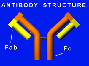
Monoclonal antibodies are powerful weapons which could be constructed to react with a desired antigen. When an antibody reacts with antigen, complement also comes into play and this combination of weaponry produces a powerful destructive force.