RHEUMATOID ARTHRITIS: SLIDES & ANIMATIONS

PAGE 2

 
Inflammation, Rheumatoid Arthritis, and the Biologics: Page 2
   

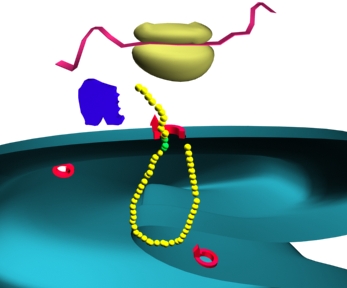
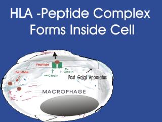
     Immunity can be broken down into two broad categories, i.e. the innate or natural immunity, and specific immunity. Innate immunity is the more ancient of the two in the animal kingdom and consists of physical barriers, nonspecific phagocytosis, and macrophage derived cytokines, such as interferon and tumor necrosis factor (TNF). Specific immunity is more recently evolved and includes T-cell cellular responses and the B cells with their antibody (humoral) responses. Slides showing antigen processing by a macrophage and subsequent presentation to a T cell will be shown. Antigen is broken down into small peptides and combined with HLA (human leukocyte antigen) molecules within the cell.

 
bacteria1.jpg (10444 bytes)   Bacteria2 in Macrophage HLA.jpg (12854 bytes)   Bacteria3 in Macrophage HLA.jpg (13618 bytes)  
Slide 9: "Antigen Processing within a macrophage is demonstrated." 32o X 240 pixels jpeg 11kb
Slide 10: "A HLA Peptide Complex forms inside the cell." 32o X 240 pixels jpeg 13kb
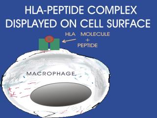
Slide 11: "The HLA Peptide Complex is displayed on the cell surface."32o X 240 pixels jpeg 14kb  


Bacteria4 in Macrophage HLA.jpg (10546 bytes)   Bacteria5 in Macrophage HLA.jpg (10699 bytes)
Windows Media Player

Real Media Player


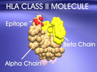
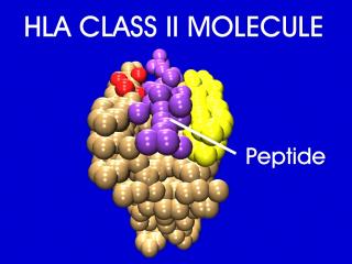
Animation 5: HLA molecule's disease causing epitope
: A 3D model of the MHC Class II molecule twirls, with the disease causing epitope designated in red.
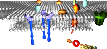

Animation 5: HLA molecule's disease causing epitope : A 3D model of theMHC Class II molecule twirls, with the disease causing epitope designated in red.

Flash Player

QuickTime Media Player

Animation 5: HLA molecules disease causing epitope
: A 3D model of the MHC Class II molecule twirls, with the disease causing epitope designated in red.


A 3D model of the MHC Class II molecule twirls, with the disease causing epitope designated in red.

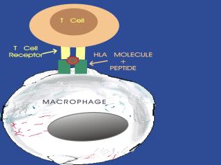
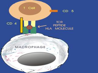
Slide 12: "Antigen is presented by the macrophage to a T cell."32o X 240 pixels jpeg 11kb    Slide 13: "Antigen is presented by the macrophage to a T cell. The CD 5 and CD4 molecules are also shown. "32o X 240 pixels jpeg 11kb
   

     The peptide-HLA (also called peptide-MHC for major histocompatibility) complex is then displayed on the surface of the antigen presenting cell. HLA molecules play a critical role in the immune response to foreign antigens and are highly polymorphic. The MHC is the genetic cornerstone of the immune response. If a peptide cannot be bound by a HLA molecule, then there will be no immune response to it. Also, it is the HLA molecule type that predisposes to autoimmune disease.


Ag binding cleft.jpg (12969 bytes)

  HLA from above.jpg (14060 bytes)   HLA c epitope.jpg (13884 bytes)  
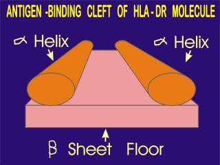
Slide 13: ""The antigen binding cleft of a HLA-DR molecule is shown."32o X 240 pixels jpeg 13kb
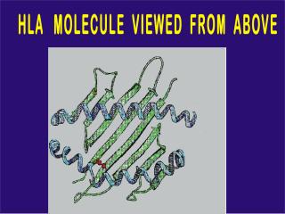
Slide 14: ""The cleft of the antigen binding site is viewed from above. "32o X 240 pixels jpeg 14kb
Slide 15: ""The are of the HLA molecule responsible for disease susceptibility is shown in red (the epitope)."32o X 240 pixels jpeg 14kb
 
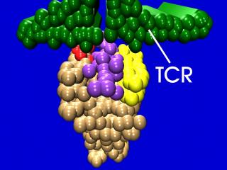
     The area of the HLA molecule responsible for disease susceptibility is diagramed in red as an epitope. The epitope’s spacial relationship to both the groove and TCR is stressed. The antigen binding cleft is shown. We will concentrate today on HLA type II molecules, which deal with exogenous antigens.
 
HLA peptide and TCR.jpg (12921 bytes) HLA pep and TCRgreen2.jpg (13593 bytes)  
 
 
Slide 16: ""The peptide is shown in purple in the antigen binding site. "32o X 240 pixels jpeg 12kb  

Slide 17: ""The trimolecular complex is shown."32o X 240 pixels jpeg 14kb

 
About Us | Contact Us | ©2005   This web site has been developed and maintained by Craig W. Wiesenhutter, M.D.   Last updated 1/03/05