OSTEOARTHRITIS: SLIDES & ANIMATIONS

PAGE 1


     This is a multimedia talk that was given in 1998 on Osteoarthritis and NSAIDs. I've left most of the original material, but I've added a great deal as well and I now include most of the animations in modern file formats i.e. flash, window media player, quickTime, and real media player.

 
  oa title.jpg (5104 bytes)    
Slide 1: "Clinic Space Ship" On my way to give a talk. 280 x 210 pixels 12kb The spaceship was a model included in an older 3dStudio max version.
Slide 2: "Osteoarthritis Title " 320 x 240 pixels 5kb drawn freehand in 3dStudio Max.

Slide 3: "Rheumatoid Arthritis Title" 300 x 225 pixels 18kb drawn freehand in 3dStudio Max.
Click Picture or here for larger slides (800 x 600 pixels) 61kb and (1024 x 768 pixels) 88kb   Click Picture or here for larger slides (800 x 600 pixels) 143kb
 
nsaid war title.jpg (7420 bytes)  
Windows Media Player
 
QuickTime Media Player
 
Flash Player


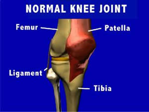
Animation 1: Normal Knee Anatomy
: 3d view of the knee anatomy.
Animation 2: Joint Stability : Joint Stability is discussed.


Animation 1: Normal Knee Anatomy
: 3d view of the knee anatomy.
Animation 2: Joint Stability : Joint Stability is discussed.

3d view of the knee anatomy. Joint Stability is discussed.

Real Media Player


Animation 1: Normal Knee Anatomy : 3d view of the knee anatomy.
.Animation 2: Joint Stability : Joint Stability is discussed.

Slide 4: "NSAID Wars Title " 320 x 240 pixels 8kb Drawn freehand in 3dStudio Max.
 

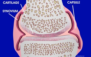
     Slide #1: In transit to Sun Valley . Slide #2 presents the title of the talk as osteoarthritis (OA). The importance of osteoarthritis is discussed. It is the most common arthritis, and it is the most common chronic illness in people over 65 years of age. There have been a lot of new advances concerning pathophysiology as well as new concepts about the development of "joint failure", which will be presented. The second part of the talk will deal with NSAIDs and will focus on side effects (Slide #4). New NSAIDs, i.e., Arthrotec, and the highly selective Cox 2 agents will be discussed. Slide #5 is a schematic drawing of a synovial joint. The structure and function of the capsule, synovium, hyaline cartilage, and subchondral bone are discussed. Slide #6 is a 3D model of a normal knee joint, anterior oblique view.  
     Joint stability stems from the congruity of the opposing structures, as well as the support given by the fibrous capsule, ligaments Slide 37, and in some cases, menisci. The chemical nature of synovial fluid also leads to an inhibition of distraction of the bones, acting as an adhesive, and offering a stabilizing force to the joint. However, muscle contraction is by far and away the most important factor in both joint stabilization and the distribution of force across the joint.
   
E-Topics: Osteoarthritis
Normal Joints: Structure and Function
 
Osteoarthritis
       "joint configuration "
       "joint lubrication "
       "joint stability "
       "normal joint defined "
       "normal knee anatomy"
       "structure & function"
       "types of joints "    

       "oa animated schematic"
       "oa clinical manifestations"
       "oa defined
       "oa gross anatomy "
       "oa joint pattern "
       "oa pathophysiology"
       "oa spinal "
       "oa summary "
       "oa typical changes "

Cartilage: Structure and Function
      "cartilage: schematic"       "cartilage: hyaline "
      "cartilage: remodeling "
      "chondrocyte"
      "collagen"

 
Flash E-books: Osteoarthritis

"Normal Joint Structure and Function and Osteoarthritis "
The original CDrom content written in 2000 for the medical students as a handout.   All of this content is also contained in the "Rheumatology Overview" E-book.

"Rheumatology Overview: Images and Animation in Arthritis and Rheumatism"
The lead lecture for the ucla second year medical school pathophysiology of disease course, covering numerous topics in Rheumatology.

Slide 5: "A schematic of a normal synovial joint. " 400 x 250 pixels 37kb Drawn freehand in CorelDraw
Slide 6: "Normal Knee Joint " 300 x 225 pixels 10kb Viewpoint Model Worked in 3dStudio Max.
Click Picture or here for larger slides (800 x 500 ) 98b  


Slide 7: Normal Knee Anatomy: Cruciate Ligaments " 300 x 225 pixels 17kb Viewpoint Model Worked in 3dStudio Max.
Slide 8: "Schematic Joint with Synovial Fluid " 300 x 225 pixels 32kb freehand 3dStudio Max

Slide 9: "Ankle with Bursal Sac Illustrated and Bitmapped Muscles and Tendons" 300 x 225 pixels 19kb Bitmaps drawn in Corel Draw. Viewpoint Model Worked in 3dStudio Max
Click Picture or here for larger slides (800 x 500 ) 98b Click Picture or here for larger slides (800 x 600 ) 121kb Click Picture or here for larger slides (800 x 600 ) 73kb
 
About Us | Contact Us | ©2005   This web site has been developed and maintained by Craig W. Wiesenhutter, M.D.   Last updated 1/21/05